
 By snap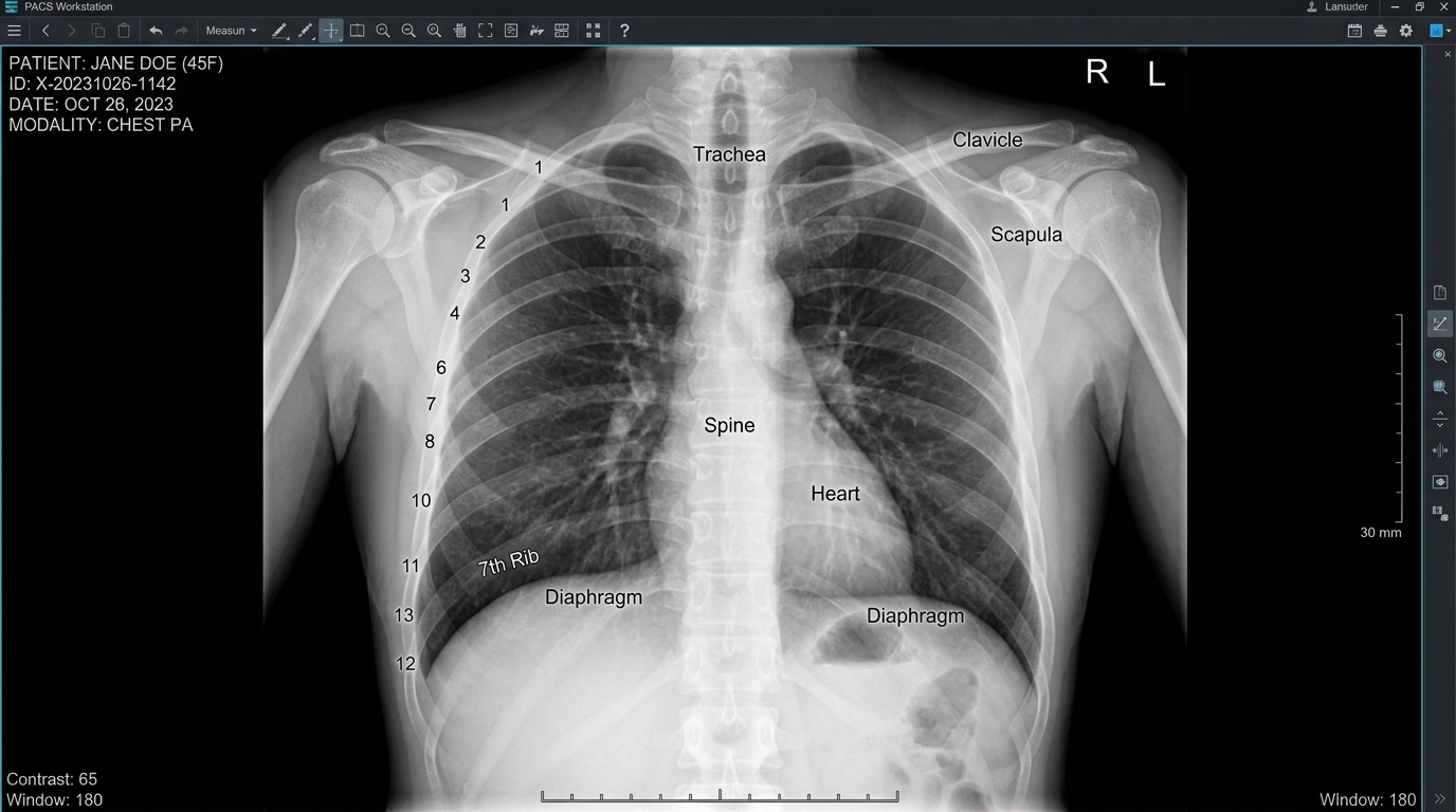
Modern medicine relies heavily on diag image technology to examine what lies beneath the skin without invasive surgery. Doctors, radiologists, and specialists use these tools daily to detect fractures, tumors, infections, and organ abnormalities. Consequently, patients receive faster diagnoses and more accurate treatment plans than ever before. This article explores the major imaging technologies available today, explains how each one works, and highlights their specific clinical applications. Furthermore, we will examine emerging trends shaping the future of internal body visualization.
Why Internal Body Imaging Matters
Physicians cannot always determine what is happening inside a patient’s body through physical examination alone. Therefore, imaging technology bridges this gap by producing detailed pictures of bones, organs, and soft tissues. Without these tools, many conditions would remain undetected until symptoms became severe or life-threatening. As a result, early detection through imaging often improves treatment outcomes significantly. Additionally, these technologies help surgeons plan procedures with greater precision and reduced risk. Medical imaging also plays a crucial role in monitoring disease progression over time. For instance, oncologists track tumor size and response to chemotherapy through repeated scans.
X-Ray Technology: The Foundation of Medical Imaging
X-rays represent the oldest and most widely used imaging method in modern healthcare facilities. This technology uses electromagnetic radiation to capture images of dense structures like bones. Consequently, doctors frequently use X-rays to diagnose fractures, dislocations, and skeletal abnormalities quickly. The process works because bones absorb radiation differently than surrounding soft tissue does. As a result, dense areas appear white on the resulting image, while softer tissue appears darker. Chest X-rays also help identify lung infections, fluid buildup, and certain cardiac conditions effectively. Despite newer technologies emerging, X-rays remain essential because they are affordable, fast, and widely accessible. However, this method has limitations when examining soft tissue or complex internal structures. Therefore, doctors often combine X-rays with other imaging methods for comprehensive diagnostic accuracy.
Computed Tomography Scans for Detailed Cross-Sections
Computed tomography, commonly known as CT scanning, offers far more detail than standard X-ray imaging. This technology combines multiple X-ray images taken from different angles around the body. Subsequently, a computer processes these images into detailed cross-sectional views of internal structures. As a result, doctors can examine organs, blood vessels, and tissues with remarkable clarity and precision. CT scans prove particularly valuable when diagnosing internal bleeding, tumors, or complex fractures after trauma. Emergency departments rely heavily on this technology because scans can be completed within minutes. Moreover, CT imaging helps guide biopsy procedures by pinpointing the exact location of abnormal tissue. Radiologists also use contrast dye during certain scans to enhance visibility of blood vessels and organs. Although CT scans expose patients to more radiation than X-rays, the diagnostic benefits often outweigh this risk. Consequently, physicians carefully weigh necessity against exposure when recommending this imaging option for patients.
Magnetic Resonance Imaging and Its Clinical Applications
Magnetic resonance imaging, or MRI, uses powerful magnets and radio waves instead of radiation. This technology excels at capturing detailed images of soft tissues, including muscles, ligaments, and organs. Therefore, MRI scans are particularly useful for diagnosing brain, spinal cord, and joint conditions. Unlike CT scans, MRI does not expose patients to ionizing radiation during the procedure. As a result, doctors often prefer this method when repeated imaging is necessary over time. Neurologists frequently order MRI scans to detect strokes, tumors, and multiple sclerosis lesions accurately. Additionally, orthopedic specialists use MRI to evaluate torn ligaments, cartilage damage, and other joint injuries. The procedure typically takes longer than CT scanning, often lasting between thirty and sixty minutes. Despite this extended duration, the exceptional image quality justifies the additional time investment for many diagnoses. However, patients with certain metal implants or pacemakers may not qualify for MRI procedures. Consequently, doctors must review patient history carefully before recommending this particular imaging approach.
Ultrasound Imaging for Real-Time Visualization
Ultrasound technology uses high-frequency sound waves to create real-time images of internal body structures. This method proves especially valuable during pregnancy monitoring, allowing doctors to observe fetal development safely. Furthermore, ultrasound helps diagnose conditions affecting the heart, liver, kidneys, and reproductive organs. Unlike other imaging methods, ultrasound produces no radiation exposure, making it safe for repeated use. As a result, doctors often choose this technology when monitoring conditions over extended periods. Cardiologists frequently use echocardiograms, a specialized ultrasound, to examine heart function and structure. Additionally, this imaging method guides needle placement during biopsies and other minimally invasive procedures. The portable nature of ultrasound equipment also makes it valuable in emergency and bedside settings. Therefore, many hospitals now use handheld ultrasound devices for rapid diagnostic assessments. Despite these advantages, ultrasound images generally offer less detail than MRI or CT scans provide.
Nuclear Medicine and Functional Imaging Techniques
Nuclear medicine imaging differs from other methods because it evaluates organ function rather than structure alone. This technology involves injecting small amounts of radioactive material into the patient’s bloodstream. Subsequently, specialized cameras detect radiation emitted from these materials to create functional images. Positron emission tomography, known as PET scanning, represents one common nuclear medicine technique used today. Oncologists frequently use PET scans to detect cancer spread and monitor treatment effectiveness over time. Additionally, this technology helps cardiologists assess blood flow and heart muscle function accurately. Nuclear medicine imaging often combines with CT scanning to produce hybrid PET-CT images for enhanced accuracy. As a result, doctors gain both structural and functional information from a single diagnostic procedure. Although radiation exposure occurs during these scans, the doses remain carefully controlled and monitored throughout treatment.
Comparing Imaging Technologies for Different Medical Needs
Choosing the appropriate imaging technology depends heavily on the specific medical condition being investigated. For bone fractures, X-rays typically provide sufficient detail without requiring more advanced technology. However, soft tissue injuries often require MRI scanning for accurate diagnosis and treatment planning. Meanwhile, emergency trauma cases frequently benefit from the speed and detail offered by CT scanning. Pregnant patients and those requiring repeated monitoring generally benefit most from ultrasound technology instead. Cancer patients, on the other hand, often require nuclear medicine imaging to track disease progression. Consequently, physicians must consider factors like radiation exposure, cost, and diagnostic accuracy when selecting methods. Additionally, patient-specific factors such as metal implants or claustrophobia influence which imaging technology becomes appropriate. Therefore, collaborative decision-making between radiologists and referring physicians ensures patients receive optimal diagnostic care.
The Role of Artificial Intelligence in Modern Imaging
Artificial intelligence increasingly plays a significant role in analyzing diag image results across healthcare systems. Machine learning algorithms now assist radiologists by flagging potential abnormalities within scans automatically. As a result, diagnostic accuracy improves while reducing the time required for image interpretation. Furthermore, AI systems help detect subtle patterns that human eyes might occasionally overlook during review. Hospitals increasingly integrate these tools into existing workflows to support, rather than replace, radiologist expertise. Consequently, patients benefit from faster turnaround times and more consistent diagnostic accuracy across facilities. Additionally, AI-powered imaging software helps standardize interpretations across different healthcare providers and geographic locations. This technology also assists in predicting disease progression based on patterns identified in historical data. Despite these advancements, human oversight remains essential for confirming diagnoses and making treatment decisions. Therefore, artificial intelligence serves as a powerful complement to traditional radiological expertise rather than a replacement.
Patient Safety and Radiation Considerations
Radiation exposure remains a significant consideration when selecting appropriate imaging technologies for patient care. Medical professionals follow strict guidelines to minimize unnecessary radiation while maintaining diagnostic image quality. As a result, technologies like ultrasound and MRI are often preferred when radiation-free alternatives exist. However, situations requiring detailed cross-sectional views sometimes necessitate CT scanning despite radiation exposure concerns. Consequently, radiologists carefully calculate appropriate radiation doses based on patient age, weight, and condition severity. Children and pregnant women receive special consideration due to increased sensitivity to radiation exposure. Therefore, alternative imaging methods are prioritized whenever clinically appropriate for these vulnerable patient populations. Additionally, healthcare facilities maintain detailed records tracking cumulative radiation exposure for patients requiring frequent imaging. This practice helps prevent excessive radiation accumulation from multiple procedures conducted over extended periods.
Future Trends in Internal Body Visualization
Emerging technologies continue transforming how healthcare providers visualize and analyze internal body structures. Three-dimensional imaging techniques now offer more detailed and interactive views than traditional two-dimensional scans. Furthermore, portable imaging devices are becoming increasingly sophisticated, bringing diagnostic capabilities directly to patient bedsides. Researchers are also developing contrast agents that provide clearer, more targeted visualization of specific tissue types. As a result, future diagnoses may become faster, more accurate, and less invasive overall. Additionally, cloud-based imaging platforms now allow specialists worldwide to collaborate on complex diagnostic cases remotely. This connectivity proves especially valuable in rural areas lacking access to specialized radiological expertise. Virtual and augmented reality applications are also emerging as tools for surgical planning and education. Consequently, medical students and surgeons can practice procedures using realistic three-dimensional anatomical models. These innovations promise to make internal body visualization more accessible, accurate, and efficient in coming years.
Conclusion
Diag image technology has transformed modern medicine by making internal body structures visible without invasive surgery. From basic X-rays to advanced nuclear medicine techniques, each method offers unique diagnostic advantages. Consequently, healthcare providers can select the most appropriate tool based on specific patient needs. Furthermore, emerging technologies like artificial intelligence and three-dimensional imaging continue enhancing diagnostic accuracy and efficiency. As these tools evolve, patients can expect faster diagnoses, more targeted treatments, and improved health outcomes. Understanding these various imaging technologies helps patients engage more actively in their own healthcare decisions. Ultimately, continued innovation in this field promises even greater precision and accessibility in the future.